Normal Retina vs. Papilledema - Trial Exhibits Inc.
Por um escritor misterioso
Last updated 06 fevereiro 2025

This illustration compares a normal retina of the left eye and a retina with papilledema. The retina with papilledema is characterized by a bulging optic disc.
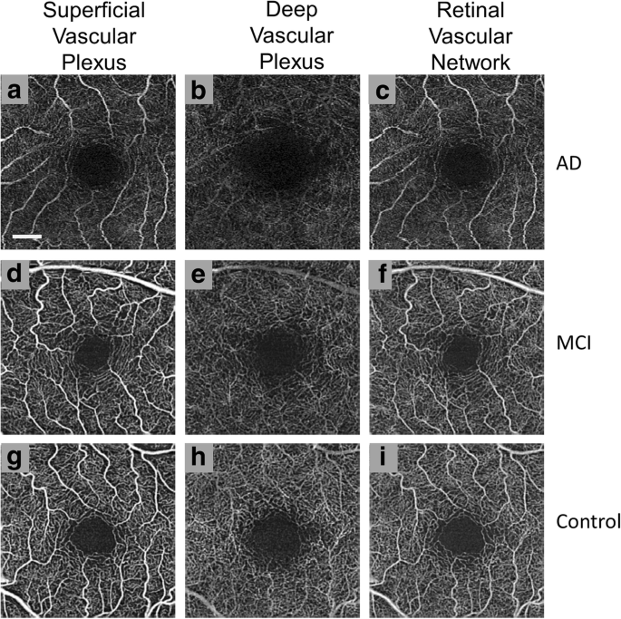
Emerging Applications of Optical Coherence Tomography Angiography (OCTA) in neurological research, Eye and Vision
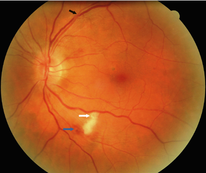
Hypertensive Fundus Changes

Spectrum of cerebrospinal fluid pressure-related ophthalmic disease.
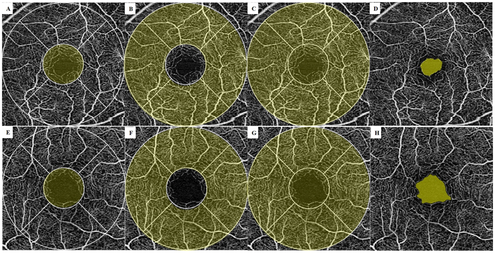
Retinal Microvascular Change in Hypertension as measured by Optical Coherence Tomography Angiography

Disorders of Optic Nerve and Visual Pathways

Optic nerve head anatomy in myopia and glaucoma, including parapapillary zones alpha, beta, gamma and delta: Histology and clinical features - ScienceDirect

Accuracy of Diagnostic Imaging Modalities for Classifying Pediatric Eyes as Papilledema Versus Pseudopapilledema - ScienceDirect
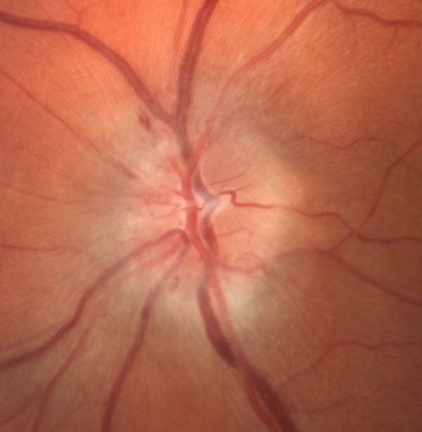
Idiopathic Intracranial Hypertension. Vision at Risk.

Photographs of the fundus showing the optic disc from the control eye
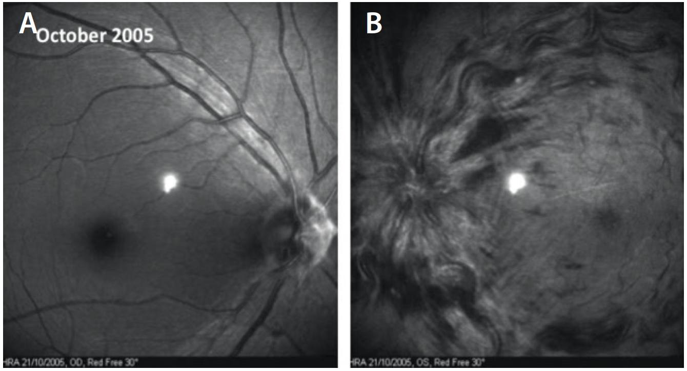
Papillophlebitis: a Closer Look - Retina Today
Recomendado para você
-
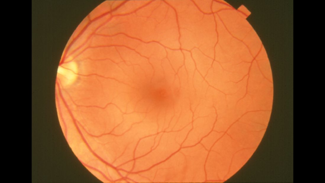 7 health problems predicted with a look into your eyes06 fevereiro 2025
 -
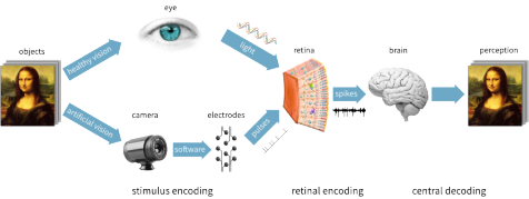 Research, Stanford Artificial Retina Project06 fevereiro 2025
 -
![Figure 1. [The normal human retina fundus]. - Webvision - NCBI](https://www.ncbi.nlm.nih.gov/books/NBK554706/bin/Archetecture_Fovea-Image006.jpg) Figure 1. [The normal human retina fundus]. - Webvision - NCBI06 fevereiro 2025
Figure 1. [The normal human retina fundus]. - Webvision - NCBI06 fevereiro 2025 -
 What Causes Retinal Detachment?06 fevereiro 2025
 -
 Descolamento de Retina: 7 Mitos Que Você Precisa Conhecer Agora06 fevereiro 2025
 -
 Retina Associates of Cleveland, Inc.06 fevereiro 2025
 -
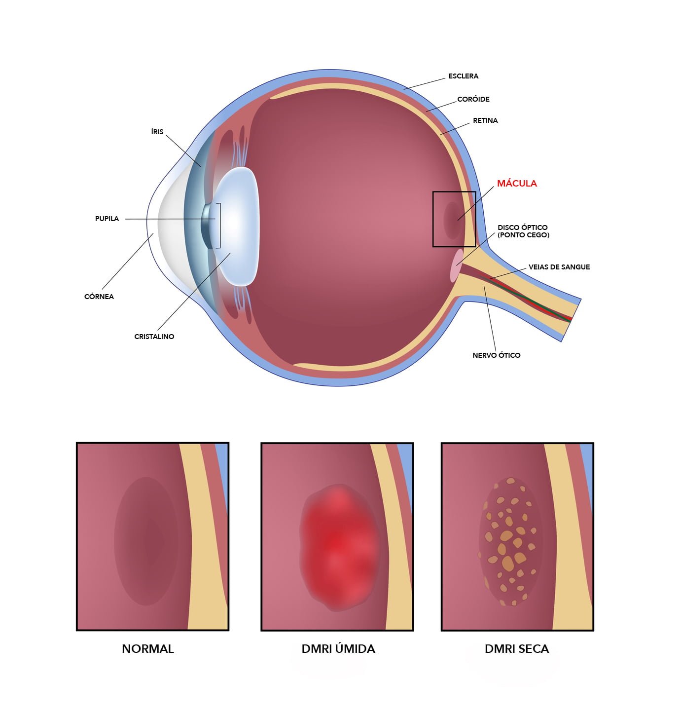 Doenças da Retina06 fevereiro 2025
 -
 Cirurgias de Retina e Vítreo Hospital de Olhos de Registro06 fevereiro 2025
 -
 Retina Imaging Village Optical06 fevereiro 2025
 -
 51,730 Retina Images, Stock Photos, 3D objects, & Vectors06 fevereiro 2025
você pode gostar
-
 oGol on X: Dois espanhóis, três italianos, três ingleses, dois portugueses, um holandês e um alemão: é assim o ranking de maiores vencedores da história da Liga dos Campeões. / X06 fevereiro 2025
 -
 Crunchyroll Adds Re:Zero Season 2 To Summer 2020 Simulcasts - Anime Herald06 fevereiro 2025
 -
 Atum - JoJo's Bizarre Encyclopedia06 fevereiro 2025
 -
 OS MELHORES JOGOS DE FUTEBOL OFFLINE PARA ANDROID06 fevereiro 2025
 -
Konami Computer Entertainment Japan (video game company, Japan) - Glitchwave video games database06 fevereiro 2025
-
 Cidade das Artes - Programação - Rei Simba06 fevereiro 2025
 -
 K ALPHABET LORE REFERANCE!!!11!1??/?1? : r/alphabetfriends06 fevereiro 2025
 -
 FIFA Mobile on iPhone 6 Plus ios 12.5.506 fevereiro 2025
 -
 Capa Livro Colorir Lol Surprise + 20 folhas (Arte Digital)06 fevereiro 2025
 -
 FENDA DO BUG!, Roblox Lumber Tycoon 206 fevereiro 2025