Morphology of Leydig cells in the testes after in vivo MCP-1 treatment.
Por um escritor misterioso
Last updated 10 março 2025


Testicular macrophages are recruited during a narrow time window by fetal Sertoli cells to promote organ-specific developmental functions

Rapid Differentiation of Human Embryonic Stem Cells into Testosterone-Producing Leydig Cell-Like Cells In vitro
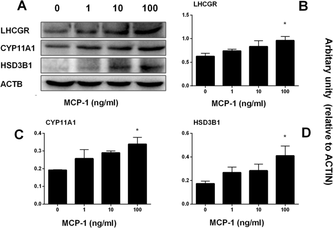
Monocyte Chemoattractant Protein-1 stimulates the differentiation of rat stem and progenitor Leydig cells during regeneration, BMC Developmental Biology

The Sertoli cell: one hundred fifty years of beauty and plasticity - França - 2016 - Andrology - Wiley Online Library

Transcription factor Dmrt1 triggers the SPRY1-NF-κB pathway to maintain testicular immune homeostasis and male fertility

Morphology of Leydig cells in the testes after in vivo PTHrP

PDF) Monocyte Chemoattractant Protein-1 stimulates the differentiation of rat stem and progenitor Leydig cells during regeneration
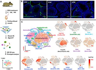
Frontiers Identification of Rat Testicular Leydig Precursor Cells by Single-Cell-RNA-Sequence Analysis

Testicular inflammation and infertility: Could chlamydial infections be contributing? - Bryan - 2020 - American Journal of Reproductive Immunology - Wiley Online Library
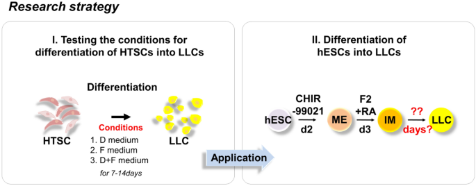
Rapid Differentiation of Human Embryonic Stem Cells into Testosterone-Producing Leydig Cell-Like Cells In vitro
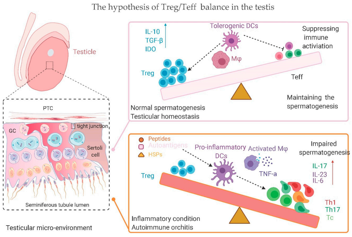
IJMS, Free Full-Text
Recomendado para você
-
 Resultado Teste Vivo Fibra Tv - Vale a Pena?10 março 2025
 -
 Vivo X100 ganha teste com Dimensity 9300 superando Snapdragon 8 Gen 3 - Canaltech10 março 2025
 -
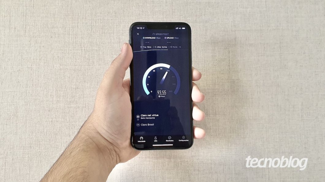 Brasil tem 74ª internet móvel mais rápida do mundo; Claro e Vivo lideram – Tecnoblog10 março 2025
 -
 RTP do Patch 2.4 de Diablo II: Resurrected, Teste Competitivo10 março 2025
 -
 Histomorphometrical evaluation of zebrafish testes after in vivo10 março 2025
 -
/i.s3.glbimg.com/v1/AUTH_08fbf48bc0524877943fe86e43087e7a/internal_photos/bs/2021/n/k/dg0dIJToA5v0aLmvm6vw/2015-11-19-batepapo41.jpg) Como testar transmissão ao vivo com um evento programado no10 março 2025
Como testar transmissão ao vivo com um evento programado no10 março 2025 -
 Vivo X100 ganha imagens oficiais, amostras de fotos e testes de desempenho - Canaltech10 março 2025
 -
 Teste Padrão Vivo Da Pele Do Crocodilo Do Corpo Vivo Para O Fundo10 março 2025
 -
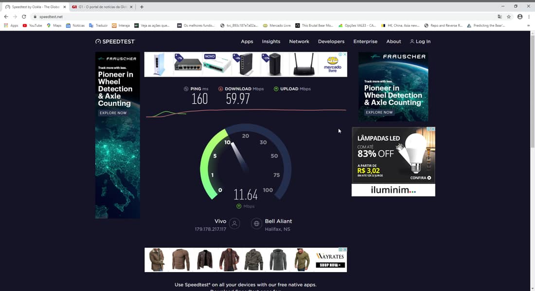 Vivo Fibra é bom? Teste do plano 300mbps/150mbps conectando a10 março 2025
 -
![Microsoft Azure DevOps - Foco em Testes Ágeis [Ao Vivo + On Demand] - Iterasys](https://cdn.eveclass.com/p/61cb81d816aa58f336ffe148/files/gallery/image/9e600cc0-fe01-11ec-a7d4-611f0e6ccdc5/thumbnail.jpg) Microsoft Azure DevOps - Foco em Testes Ágeis [Ao Vivo + On Demand] - Iterasys10 março 2025
Microsoft Azure DevOps - Foco em Testes Ágeis [Ao Vivo + On Demand] - Iterasys10 março 2025
você pode gostar
-
 Planetshakers' Youth Band planetboom Releases 'Greatest In The World' Double-Single10 março 2025
 -
 How to Use Now Playing in Google Pixel 8 and Pixel 8 Pro10 março 2025
 -
Hank Hill - Interesting idea. I like that Tom Hanks fella!10 março 2025
-
 Student Testimonials10 março 2025
 -
![PSA] Remember the mod developer makes mods on their spare time](https://external-preview.redd.it/29hmvsXPsp9fk9LuGxYVlzsXg21WPmKsI_QMpuNFr4k.png?width=640&crop=smart&auto=webp&s=036726cc46ddfa99bfdbf33aa60577ae11598619) PSA] Remember the mod developer makes mods on their spare time10 março 2025
PSA] Remember the mod developer makes mods on their spare time10 março 2025 -
 boruto dublado em portugues ep 5310 março 2025
 -
 My Singing Monsters Collections 202310 março 2025
 -
Ver episódios de Kono Yo no Hate de Koi wo Utau Shoujo YU-NO em streaming10 março 2025
-
 Assistir Kanojo, Okarishimasu 2 Episódio 12 » Anime TV Online10 março 2025
 -
Russian Alphabet Lore Humanized Part 1010 março 2025

