Topographic sketch. Coronal view of bilateral DRT (orange). Patient
Por um escritor misterioso
Last updated 26 fevereiro 2025


Structure of Long-Range Direct and Indirect Spinocerebellar Pathways as Well as Local Spinal Circuits Mediating Proprioception

Crossing nerve transfer drives sensory input–dependent plasticity for motor recovery after brain injury

DTI for brain targeting: Diffusion weighted imaging fiber tractography—Assisted deep brain stimulation - ScienceDirect
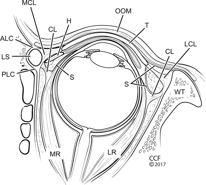
Applied Surgical Anatomy of the Ocular Adnexa

Is Graphene Shortening the Path toward Spinal Cord Regeneration?

Visual signs and symptoms of multiple system atrophy - Armstrong - 2014 - Clinical and Experimental Optometry - Wiley Online Library

Journal of Comparative Neurology, Systems Neuroscience Journal

DTI for brain targeting: Diffusion weighted imaging fiber tractography—Assisted deep brain stimulation - ScienceDirect
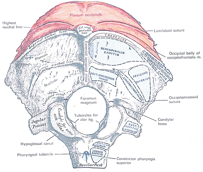
The Neck: Development and Evolution
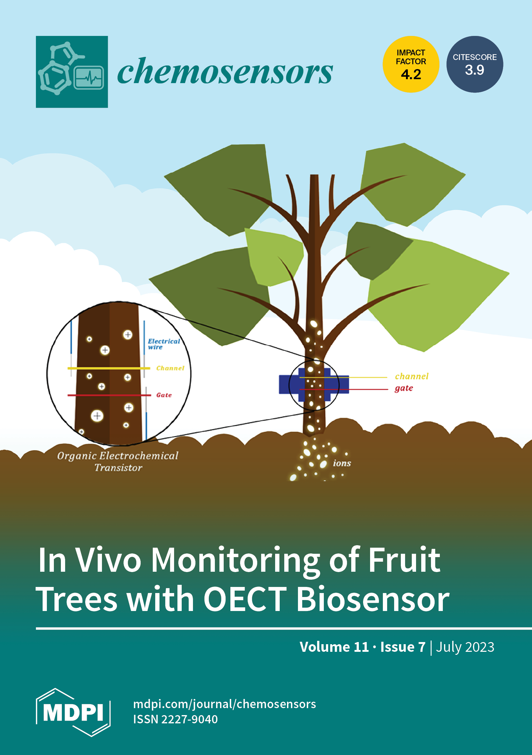
Chemosensors July 2023 - Browse Articles

DTI for brain targeting: Diffusion weighted imaging fiber tractography—Assisted deep brain stimulation - ScienceDirect
Recomendado para você
-
 SCP-PL-007 - SCP International26 fevereiro 2025
 -
 SCP-006-PT - Fundação SCP26 fevereiro 2025
 -
 SCP APK for Android Download26 fevereiro 2025
 -
 Cypher 007 Mobile Game Launches26 fevereiro 2025
 -
 CapCut_scp 007 asli26 fevereiro 2025
 -
 Tapete Nitro Concepts Sporting Clube de Portugal, Fan Editi26 fevereiro 2025
 -
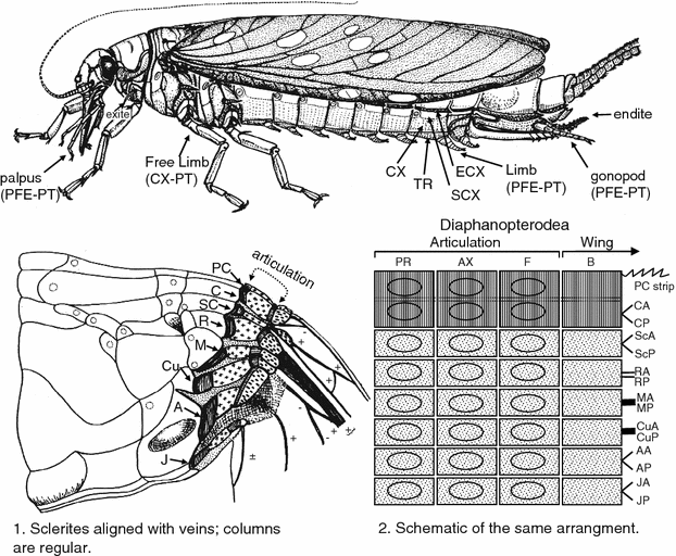 Phylogeny of Higher Taxa in Insecta: Finding Synapomorphies in the Extant Fauna and Separating Them from Homoplasies26 fevereiro 2025
 -
 Cortante Flower Burst Spellbinders para Sizzix S2-00726 fevereiro 2025
 -
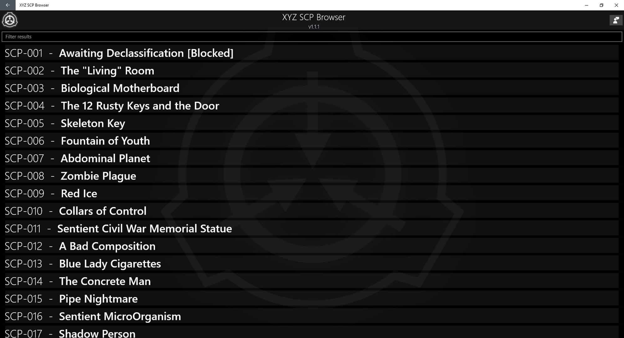
XYZ SCP Browser - Microsoft Apps26 fevereiro 2025
-
 SCP-034 Yugioh card26 fevereiro 2025
você pode gostar
-
 Assistir Chainsaw Man Episódio 11 Online - Animes BR26 fevereiro 2025
 -
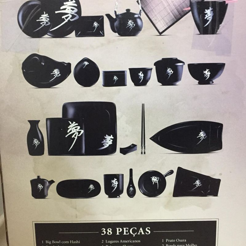
Jogo de Jantar Japones Coleção Sonho Oriental Caras 38 Peças Nunca Usado, Móvel de Cozinha Camicado Nunca Usado 3556868926 fevereiro 2025
-
 How to play music in your Roblox games - Pro Game Guides26 fevereiro 2025
 -
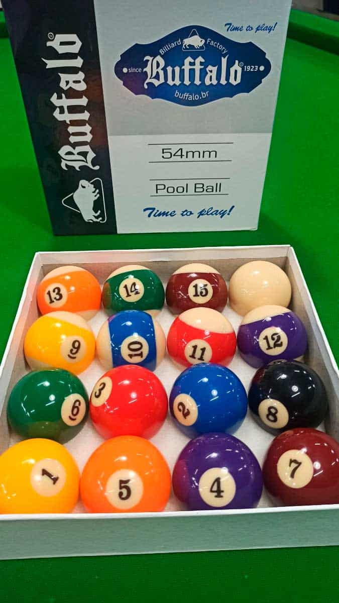 Jogo de Bola Buffalo numerada faixada (com 16 bolas) - 54 mm26 fevereiro 2025
 -
 File:Gears 5 Hivebuster Squad.png - Wikipedia26 fevereiro 2025
 -
 Conjunto de cactos em estilo simples de desenho animado isolado no26 fevereiro 2025
 -
 How to Pronounce Oof? (CORRECTLY)26 fevereiro 2025
 -
 Wild Horse Islands Codes – News Codes! – Gamezebo26 fevereiro 2025
 -
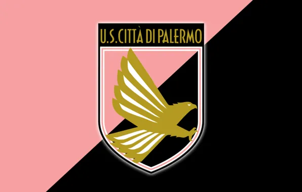 Wallpaper football club, Series A, Palermo, Palermo, Pink-black26 fevereiro 2025
 -
kasha new gods nezha reborn|Pesquisa do TikTok26 fevereiro 2025