PDF] Brain Tumor Segmentation of MRI Images Using Processed Image Driven U-Net Architecture
Por um escritor misterioso
Last updated 01 março 2025
![PDF] Brain Tumor Segmentation of MRI Images Using Processed Image Driven U-Net Architecture](https://d3i71xaburhd42.cloudfront.net/c750894747d2b3f841de55922b2b68794295de27/7-Table3-1.png)
A fully automatic methodology to handle the task of segmentation of gliomas in pre-operative MRI scans is developed using a U-Net-based deep learning model that reached high-performance accuracy on the BraTS 2018 training, validation, as well as testing dataset. Brain tumor segmentation seeks to separate healthy tissue from tumorous regions. This is an essential step in diagnosis and treatment planning to maximize the likelihood of successful treatment. Magnetic resonance imaging (MRI) provides detailed information about brain tumor anatomy, making it an important tool for effective diagnosis which is requisite to replace the existing manual detection system where patients rely on the skills and expertise of a human. In order to solve this problem, a brain tumor segmentation & detection system is proposed where experiments are tested on the collected BraTS 2018 dataset. This dataset contains four different MRI modalities for each patient as T1, T2, T1Gd, and FLAIR, and as an outcome, a segmented image and ground truth of tumor segmentation, i.e., class label, is provided. A fully automatic methodology to handle the task of segmentation of gliomas in pre-operative MRI scans is developed using a U-Net-based deep learning model. The first step is to transform input image data, which is further processed through various techniques—subset division, narrow object region, category brain slicing, watershed algorithm, and feature scaling was done. All these steps are implied before entering data into the U-Net Deep learning model. The U-Net Deep learning model is used to perform pixel label segmentation on the segment tumor region. The algorithm reached high-performance accuracy on the BraTS 2018 training, validation, as well as testing dataset. The proposed model achieved a dice coefficient of 0.9815, 0.9844, 0.9804, and 0.9954 on the testing dataset for sets HGG-1, HGG-2, HGG-3, and LGG-1, respectively.
![PDF] Brain Tumor Segmentation of MRI Images Using Processed Image Driven U-Net Architecture](https://www.mdpi.com/computers/computers-10-00139/article_deploy/html/images/computers-10-00139-g003.png)
Computers, Free Full-Text
![PDF] Brain Tumor Segmentation of MRI Images Using Processed Image Driven U-Net Architecture](https://onlinelibrary.wiley.com/cms/asset/85e7cbd1-a542-4c37-9d7f-16c756eeef98/ima22571-fig-0008-m.jpg)
International Journal of Imaging Systems and Technology, IMA
![PDF] Brain Tumor Segmentation of MRI Images Using Processed Image Driven U-Net Architecture](https://www.researchgate.net/publication/349902492/figure/fig1/AS:1024432529215497@1621255156793/Proposed-tumor-segmentation-and-classification-architecture.png)
Proposed tumor segmentation and classification architecture
![PDF] Brain Tumor Segmentation of MRI Images Using Processed Image Driven U-Net Architecture](https://media.springernature.com/lw685/springer-static/image/art%3A10.1186%2Fs13244-020-00869-4/MediaObjects/13244_2020_869_Fig5_HTML.png)
Convolutional neural networks for brain tumour segmentation, Insights into Imaging
![PDF] Brain Tumor Segmentation of MRI Images Using Processed Image Driven U-Net Architecture](https://file.techscience.com/ueditor/files/cmes/TSP_CMES_128-2/TSP_CMES_14107/TSP_CMES_14107/Images/CMES_14107-fig-5.png/mobile_webp)
MRI Brain Tumor Segmentation Using 3D U-Net with Dense Encoder Blocks and Residual Decoder Blocks
![PDF] Brain Tumor Segmentation of MRI Images Using Processed Image Driven U-Net Architecture](https://media.springernature.com/lw685/springer-static/image/art%3A10.1038%2Fs41598-021-90428-8/MediaObjects/41598_2021_90428_Fig12_HTML.jpg)
Brain tumor segmentation based on deep learning and an attention mechanism using MRI multi-modalities brain images
![PDF] Brain Tumor Segmentation of MRI Images Using Processed Image Driven U-Net Architecture](https://media.springernature.com/m685/springer-static/image/art%3A10.1038%2Fs41598-023-47107-7/MediaObjects/41598_2023_47107_Fig1_HTML.png)
Utilizing deep learning via the 3D U-net neural network for the delineation of brain stroke lesions in MRI image
![PDF] Brain Tumor Segmentation of MRI Images Using Processed Image Driven U-Net Architecture](https://www.sciltp.com/journals/public/site/images/ijndi/pic/173-3.jpg)
Deep Learning Attention Mechanism in Medical Image Analysis: Basics and Beyonds-Scilight
![PDF] Brain Tumor Segmentation of MRI Images Using Processed Image Driven U-Net Architecture](https://ietresearch.onlinelibrary.wiley.com/cms/asset/783f8011-bdec-422a-a43e-d006814a5ad8/ipr212219-fig-0002-m.jpg)
Brain tumour cell segmentation and detection using deep learning networks - Bagyaraj - 2021 - IET Image Processing - Wiley Online Library
![PDF] Brain Tumor Segmentation of MRI Images Using Processed Image Driven U-Net Architecture](https://media.licdn.com/dms/image/D5612AQHmXP7oVdF7lg/article-cover_image-shrink_600_2000/0/1698339241926?e=2147483647&v=beta&t=KD2ddAt0ARBrWkFWt9qFoICdB7mtpNzg5iOUr3HikdQ)
How U and W Net Architecture in Computer Vision shaped some real work problems in Medical
![PDF] Brain Tumor Segmentation of MRI Images Using Processed Image Driven U-Net Architecture](https://www.degruyter.com/document/doi/10.1515/jisys-2022-0206/asset/graphic/j_jisys-2022-0206_fig_003.jpg)
A novel deep learning-based brain tumor detection using the Bagging ensemble with K-nearest neighbor
GitHub - IAmSuyogJadhav/3d-mri-brain-tumor-segmentation-using-autoencoder-regularization: Keras implementation of the paper 3D MRI brain tumor segmentation using autoencoder regularization by Myronenko A. (
![PDF] Brain Tumor Segmentation of MRI Images Using Processed Image Driven U-Net Architecture](https://production-media.paperswithcode.com/tasks/brain-tumor-segmentation_Omf9jBU.png)
Brain Tumor Segmentation
![PDF] Brain Tumor Segmentation of MRI Images Using Processed Image Driven U-Net Architecture](https://www.mdpi.com/computers/computers-10-00139/article_deploy/html/images/computers-10-00139-g004.png)
Computers, Free Full-Text
Recomendado para você
-
Brain test level 191 - Cuaca hujan membuatku muram #jawaban #Games #br01 março 2025
-
 Solutions Brain Test Niveau 191 à 20001 março 2025
 -
 what is level 191 on brain test|TikTok Search01 março 2025
 -
![Forum Game: Volume the 6th - Count to 20 before a Mod/Staff post 0! Wins - 126 [Highest number in this thread: 191] (Part 1) - Community Content - Empires & Puzzles Community Forum](https://global.discourse-cdn.com/smallgiantgames/original/3X/3/9/39484c75491a682890546bf8a8bf64277cd9c3e4.jpeg) Forum Game: Volume the 6th - Count to 20 before a Mod/Staff post 0! Wins - 126 [Highest number in this thread: 191] (Part 1) - Community Content - Empires & Puzzles Community Forum01 março 2025
Forum Game: Volume the 6th - Count to 20 before a Mod/Staff post 0! Wins - 126 [Highest number in this thread: 191] (Part 1) - Community Content - Empires & Puzzles Community Forum01 março 2025 -
 Thank you u/Bogi_D for signing my #1. I'm a cage person now! : r/CollectibleAvatars01 março 2025
 -
 Behold and See 4: Human Anatomy and Health: Samples01 março 2025
 -
brain test nivel 113 - 114 - 115 - 116 - 117 - 118 - 119 - 120 - 121 - 122 - 123, By Brian Test Gamingdf01 março 2025
-
 Maker de Aplicativos01 março 2025
 -
 Dized Rules, Boop01 março 2025
 -
 Serika and Shiroko thought Sensei is into cleaning butts after seeing what his watching on his phone by C/H : r/BlueArchive01 março 2025


você pode gostar
-
 Goku, Vegeta, broly dbs Poster for Sale by Yashdusane01 março 2025
 -
![[21 in 1] Benazcap Accessories Kit Compatible with Nintendo Switch OLED, Travel Accessory Bundle with Carry Case Screen Protector Joy-con Grips & More : Video Games](https://m.media-amazon.com/images/W/MEDIAX_792452-T2/images/I/81SVaBXu0HL._AC_UF894,1000_QL80_.jpg) [21 in 1] Benazcap Accessories Kit Compatible with Nintendo Switch OLED, Travel Accessory Bundle with Carry Case Screen Protector Joy-con Grips & More : Video Games01 março 2025
[21 in 1] Benazcap Accessories Kit Compatible with Nintendo Switch OLED, Travel Accessory Bundle with Carry Case Screen Protector Joy-con Grips & More : Video Games01 março 2025 -
Wolfoo fanmade Uwu Aoa01 março 2025
-
 YongFoto 5x3ft Happy New Year Backdrop 2024 Golden01 março 2025
 -
 Microsoft Word 200701 março 2025
 -
 GTA 6 Vice City map's massive secrets revealed after in-depth01 março 2025
 -
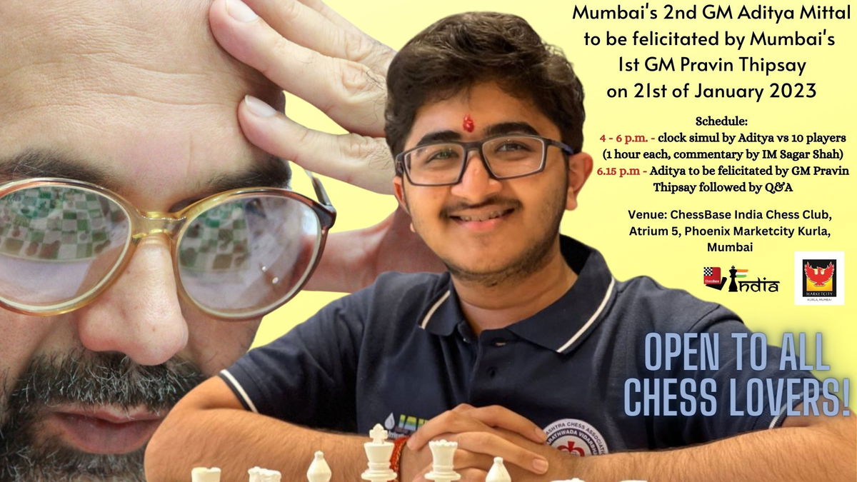 Mumbai's 2nd GM Aditya Mittal to be felicitated by Mumbai's 1st GM Pravin Thipsay - ChessBase India01 março 2025
 -
 LEAF REST: Player Home!!- Xbox Modded Skyrim Mod Showcase01 março 2025
 -
 ⚫🌟🫥M🫥🌟⚫ on X: @JamesMarshallHa You were expecting SHINY01 março 2025
 -
 Proper Jogging Form: How to Jog Like a Pro - Steel Supplements01 março 2025
